Nieskategoryzowane pliki
Z Brain-wiki
Poniżej wyświetlono co najwyżej 250 wyników w zakresie od 1051 do 1300.
Zobacz (poprzednie 250 | następne 250) (20 | 50 | 100 | 250 | 500)
 Kwrite Chaos.png 614 × 331; 13 KB
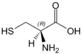 L-Cysteine(wedged bonds).png 594 × 397; 6 KB
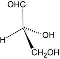 L-Glycerinaldehyd Keilstrichformel.png 500 × 495; 19 KB
 L-Tryptophan - L-Tryptophan.svg 277 × 125; 9 KB
 L-Weinsäure.svg 129 × 137; 12 KB
 L-alanine-skeletal.svg 310 × 221; 7 KB
 L-allo-Threonine.svg 191 × 126; 11 KB
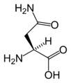 L-asparagine-skeletal.png 1267 × 1496; 23 KB
 L-aspartic-acid-skeletal.png 1267 × 1487; 26 KB
 L-leucine-skeletal.svg 212 × 223; 12 KB
 L-lysine-skeletal.png 1977 × 1462; 23 KB
 L-proline-skeletal.png 1355 × 1046; 16 KB
 L-serine-skeletal.png 1267 × 1115; 20 KB
 L-threonine-skeletal.png 1396 × 1268; 22 KB
 L-tyrosine-skeletal.png 2000 × 1643; 31 KB
 L-valine-skeletal.svg 309 × 263; 2 KB
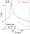 L3 z fit.png 300 × 338; 19 KB
 LA2-katalogkort.jpg 908 × 776; 67 KB
 LDA bank.003.png 810 × 647; 92 KB
 LDA bank1.png 910 × 293; 73 KB
 LDA bank2.png 798 × 653; 92 KB
 LDA bank2 ANN.png 1024 × 768; 290 KB
 LDA bank DNN.png 1024 × 768; 339 KB
 LTI rys.001.png 800 × 600; 46 KB
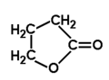 Lakton kwasu gamma-hydroksymasłowego.png 183 × 135; 651 bajtów
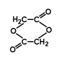 Laktyd kwasu glikolowego.png 170 × 172; 737 bajtów
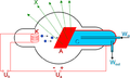 Lampa rtg.png 613 × 366; 42 KB
 Laser DSC09088.JPG 120 × 118; 4 KB
 Laurencia.jpg 900 × 722; 561 KB
 Lauterbur eksperyment 1.png 980 × 1194; 67 KB
 Lauterbur eksperyment 2.png 987 × 1194; 77 KB
 Lead electrolytic and 1cm3 cube.jpg 5260 × 3341; 3,41 MB
 Leds.png 800 × 525; 121 KB
 Leminiskata.png 556 × 274; 61 KB
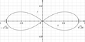
 Lemniskata.png 669 × 646; 99 KB
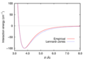 Lennard-Jones-2Ar.png 300 × 210; 20 KB
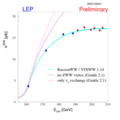 Lep ww cros2.png 300 × 300; 19 KB
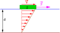 Lepkosc.png 240 × 132; 2 KB
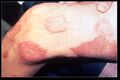 Leprosy thigh demarcated cutaneous lesions.jpg 3999 × 2670; 925 KB
 Leptin.png 928 × 726; 268 KB
 Light dispersion conceptual waves.gif 640 × 480; 1,14 MB
 Lin log reg.png 800 × 312; 112 KB
 Lina1.png 360 × 113; 3 KB
 Lina2.png 304 × 146; 5 KB
 Line pattern.png 6676 × 1226; 6 KB
 Linfit01a.gif 652 × 526; 19 KB
 Linie x ft.png 762 × 603; 1,32 MB
 Linie xy ft.png 762 × 603; 1,32 MB
 Linie y ft.png 762 × 603; 1,32 MB
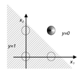
 Liniowa separowalnosc.png 763 × 347; 35 KB
 Linux-prompt.png 526 × 148; 20 KB
 Lipid bilayer section.gif 300 × 195; 124 KB
 Lipofuscin neuro.jpg 900 × 940; 217 KB
 Lista 4.svg 998 × 267; 29 KB
 Lista 4 różne.svg 998 × 267; 35 KB
 Local contrast.png 1367 × 652; 10 KB
 Lock-jaw 2857.jpg 700 × 1042; 93 KB
 Logo.jpg 1339 × 222; 71 KB
 Logo pelne KNI.png 1400 × 543; 44 KB
 Lor1.png 654 × 551; 52 KB
 Lor2.png 654 × 551; 49 KB
 Lor3.png 654 × 551; 54 KB
 Lor4.png 654 × 551; 25 KB
 Lor5.png 654 × 551; 28 KB
 Lor6.png 654 × 551; 27 KB
 Lti mtf.png 1454 × 2601; 448 KB
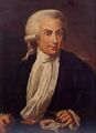 Luigi Galvani.jpg 400 × 550; 19 KB
 Lung9009f-100x.jpg 1280 × 960; 214 KB
 Lung9009f-400x.jpg 1280 × 960; 207 KB
 Lung9009f-40x.jpg 1280 × 960; 301 KB
 Lymphocyte.png 73 × 77; 4 KB
 Lymphocyte2.jpg 856 × 592; 72 KB
 M-krezol.png 330 × 167; 1 KB
 M-ksylen.png 136 × 66; 2 KB
 MEG maszyna.png 960 × 720; 979 KB
 MM 4.png 297 × 301; 3 KB
 MO2.png 313 × 256; 10 KB
 MO3.png 267 × 240; 6 KB
 MO4.png 375 × 95; 7 KB
 MP LFP 13 02.jpg 560 × 420; 37 KB
 MP rys 1 1 big.png 938 × 750; 69 KB
 MP rys 1 big.png 1117 × 871; 47 KB
 MP sym 2 1 big.png 938 × 750; 304 KB
 MP sym 2 big.png 1117 × 803; 126 KB
 MS Demyelinisation KB 10x.jpg 2080 × 1544; 3,22 MB
 MTBE-2D-skeletal.png 1100 × 792; 28 KB
 Macierz.svg 311 × 280; 21 KB
 Macrophage.jpg 1280 × 1024; 279 KB
 Macrophage.png 174 × 149; 17 KB
 Magnus effect.png 300 × 266; 29 KB
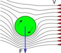 Magnus effect.svg.png 300 × 266; 29 KB
 Main biol.jpg 2198 × 1704; 2,21 MB
 Main eeg1.jpg 668 × 208; 34 KB
 Main eeg2.png 265 × 145; 60 KB
 Main math.png 420 × 181; 298 KB
 Main phys.png 302 × 263; 68 KB
 Main protein structure levels pl.svg 434 × 757; 466 KB
 Makro-edytor.png 594 × 296; 109 KB
 Malondialdehyde.png 437 × 254; 8 KB
 Malonyl.png 450 × 210; 4 KB
 Mandelbrot.png 304 × 341; 56 KB
 Mandril.jpg 800 × 1200; 1,3 MB
 Mapa.png 2000 × 1017; 173 KB
 Mapka tf erds.png 378 × 388; 108 KB
 Mapki ECoG.jpg 1024 × 768; 221 KB
 Mapowanie.png 527 × 448; 551 KB
 Mastocyty.jpg 778 × 600; 81 KB
 Maszyna pl.jpg 2500 × 2808; 1,8 MB
 MatematykaOptykaOkularowa cwicz sem1 picture 1.svg 422 × 242; 152 KB
 MatematykaOptykaOkularowa cwicz sem1 picture 10.png 212 × 215; 3 KB
 MatematykaOptykaOkularowa cwicz sem1 picture 11.png 231 × 216; 5 KB
 MatematykaOptykaOkularowa cwicz sem1 picture 12.png 234 × 220; 2 KB
 MatematykaOptykaOkularowa cwicz sem1 picture 13.png 223 × 224; 3 KB
 MatematykaOptykaOkularowa cwicz sem1 picture 14.png 171 × 174; 7 KB
 MatematykaOptykaOkularowa cwicz sem1 picture 15.png 301 × 79; 4 KB
 MatematykaOptykaOkularowa cwicz sem1 picture 16.png 320 × 79; 4 KB
 MatematykaOptykaOkularowa cwicz sem1 picture 17.png 399 × 119; 6 KB
 MatematykaOptykaOkularowa cwicz sem1 picture 5.png 158 × 158; 2 KB
 MatematykaOptykaOkularowa cwicz sem1 picture 6.png 256 × 256; 7 KB
 MatematykaOptykaOkularowa cwicz sem1 picture 7.png 197 × 199; 9 KB
 MatematykaOptykaOkularowa cwicz sem1 picture 8.png 276 × 248; 9 KB
 MatematykaOptykaOkularowa cwicz sem1 picture 9.png 199 × 196; 3 KB
 Matkula.png 568 × 495; 24 KB
 Matlab splash.png 392 × 391; 101 KB
 Matldeb1.gif 669 × 279; 12 KB
 Matldeb2.gif 667 × 278; 13 KB
 Matldeb3.gif 669 × 271; 13 KB
 Matldeb4.gif 669 × 276; 13 KB
 Matldeb5.gif 937 × 849; 40 KB
 Matrix1+2+3.png 660 × 1659; 185 KB
 Matsurf.png 568 × 495; 31 KB
 Maxwell.png 194 × 317; 4 KB
 Mbmc podstawy 1.png 726 × 380; 69 KB
 Mechanizm reakcji addycji elektrofilowej do alkenów.png 920 × 380; 2 KB
 Mechanizm reakcji eliminacji drugiego rzędu (E2).png 700 × 170; 2 KB
 Mechanizm reakcji eliminacji pierwszego rzędu (E1).png 430 × 230; 1 KB
 Mechanizm reakcji hydrolizy estrów w środowisku zasadowym.png 855 × 235; 2 KB
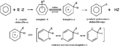 Mechanizm reakcji substytucji elektrofilowej w benzenie.png 1442 × 560; 10 KB
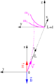
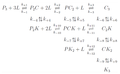 Mechanizm wiązania ligandu w procesie dwuetapowym.png 2959 × 1837; 521 KB
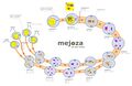 Mejoza schemat.jpg 2015 × 1318; 383 KB
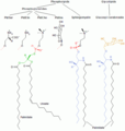 Membrane lipids.png 579 × 607; 10 KB
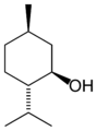 Menthol-skeletal.png 1100 × 1488; 20 KB
 Menthol skeletal.svg 347 × 436; 2 KB
 Mesenchyma.JPG 800 × 600; 70 KB
 Meso-Weinsäure Spiegel.svg 137 × 137; 12 KB
 Mesomeric peptide bond.svg 636 × 200; 24 KB
 Metanal.svg 84 × 77; 4 KB
 Methane-2D-stereo.svg 512 × 528; 5 KB
 Methionin - Methionine.svg 265 × 121; 11 KB
 Metoda parabol.png 250 × 387; 17 KB
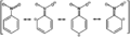 Mezomery nitrobenzenu.png 1050 × 305; 3 KB
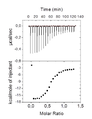 Miareczkowanie kalorymetryczne metodą ITC - przykład.png 265 × 344; 17 KB
 Michaelis-Menten.png 239 × 194; 1 KB
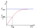
 Michaelis-Menten saturation curve of an enzyme reaction.svg 841 × 588; 20 KB
 Michealis-Menten model.pdf 0 × 0; 210 KB
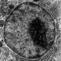 Micrograph of a cell nucleus.png 200 × 199; 33 KB
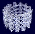 Microtuble.jpg 300 × 286; 21 KB
 Miejsca reaktywne w cząsteczce kwasu karboksylowego.png 220 × 140; 621 bajtów
 Miernik wielofunkcyjny.jpg 400 × 323; 15 KB
 Mieszanie.png 1787 × 1054; 610 KB
 Mikroglej 1.jpg 480 × 357; 20 KB
 MikroskopBresser.jpg 1343 × 596; 261 KB
 Mikroskop optyczny.jpg 2233 × 3355; 427 KB
 Milchsäure Enantiomerenpaar.svg 298 × 148; 24 KB
 Minicom.png 573 × 205; 12 KB
 Minkowski.png 286 × 256; 5 KB
 Minus exp.png 510 × 362; 19 KB
 Miofibroblasty.jpg 800 × 600; 200 KB
 Misc pollen.jpg 788 × 600; 94 KB
 Miseczkowe 1.png 450 × 277; 166 KB
 Miseczkowe 2a.png 450 × 390; 268 KB
 Miseczkowe 2b.png 450 × 390; 270 KB
 Miseczkowe 3.png 450 × 352; 207 KB
 Miseczkowe 4.png 451 × 476; 295 KB
 Miseczkowe czepek.png 450 × 216; 208 KB
 Mitochondriaa.jpg 640 × 480; 96 KB
 Mitoza schemat.jpg 1422 × 1050; 258 KB
 MięsieńSercowy1.jpg 750 × 600; 139 KB
 MięśnieGładkie1.jpg 607 × 599; 68 KB
 MięśnieSzkieletowe1.jpg 2191 × 1630; 1,24 MB
 MnistExamplesModified.png 557 × 327; 70 KB
 Moc zasady - SN2 a E2.gif 341 × 131; 2 KB
 Mod 2 drgań wahadeł sprzężonych.png 180 × 157; 9 KB
 Mod 2 drgań wahadeł sprzężonych 1.png 180 × 164; 7 KB
 ModelTNF-alfa.png 592 × 600; 215 KB
 Model LTI.png 434 × 153; 9 KB
 Model pentan i neopentan.png 600 × 400; 35 KB
- Modelowanie molekularne 2.pdf 0 × 0; 1,17 MB
 Modulacja fala prostokatna.png 1053 × 831; 56 KB
 Modulation depth.png 1367 × 652; 9 KB
 Molecular orbitals.svg 334 × 170; 139 KB
 Mono-dicom-meta.png 1235 × 510; 314 KB
 Monocyte.png 205 × 91; 18 KB
 Monosiga Brevicollis Phase.jpg 1500 × 981; 292 KB
 Morfologia-dylacja.png 654 × 551; 16 KB
 Morfologia-erozja.png 654 × 551; 16 KB
 Morfologia-otwarcie.png 654 × 551; 16 KB
 Morfologia-zakmniecie.png 654 × 551; 16 KB
 Morfologia-zaszumiony1.png 654 × 551; 19 KB
 Morfologia-zaszumiony2.png 654 × 551; 16 KB
 Morfologia-zaszumiony3.png 654 × 551; 16 KB
 Morfologia-zaszumiony4.png 654 × 551; 16 KB
 Morfologia1.png 654 × 551; 16 KB
 Mostek.png 180 × 181; 5 KB
 MotoAB.png 621 × 231; 4 KB
 Moto pot.png 1332 × 936; 379 KB
 Motor.png 821 × 615; 196 KB
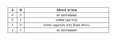 Motor tabela.png 621 × 221; 8 KB
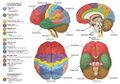 Mozg.jpeg 1024 × 715; 247 KB
- Mp5 help.pdf 0 × 0; 972 KB
 Mri basic.png 386 × 480; 6 KB
 Mrowczan etylu.svg 324 × 211; 9 KB
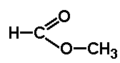 Mrówczan metylu.png 168 × 88; 439 bajtów
 Mtf kontrast.png 1027 × 616; 5 KB
 Mu niejednorodne.png 543 × 105; 7 KB
 Multi1.gif 660 × 553; 48 KB
 My data.txt ; 49 bajtów
 N,N-dimetylo-izopropyloamina.png 105 × 53; 1 KB
 N-alkiloacetamid.svg 258 × 218; 5 KB
 N-metyloanilina.png 148 × 225; 833 bajtów
 NADP+ phys.svg 363 × 608; 40 KB
 NAND.png 217 × 189; 9 KB
 NAT.png 879 × 490; 116 KB
 NN acetamid.svg 323 × 331; 6 KB
 Nablonek2.jpg 2048 × 1536; 625 KB
 NablonekPsa1.jpg 800 × 600; 184 KB
 NabłonekMigawkowy-UW1-40x.jpg 1024 × 768; 60 KB
 NabłonekMigawkowy-UW2-40x.jpg 1024 × 768; 59 KB
 NabłonekMigawkowy-UW3-100x.jpg 1024 × 768; 100 KB
 NabłonekMigawkowy-UW4-100x.jpg 1024 × 768; 111 KB
 NabłonekMigawkowy-UW5-400x.jpg 1024 × 768; 102 KB
 NabłonekMigawkowy-UW6-400x.jpg 1024 × 768; 102 KB
 NabłonekPłaski-UW1-40x.jpg 1024 × 768; 210 KB
 NabłonekPłaski-UW2-40x.jpg 1024 × 768; 188 KB
 NabłonekPłaski-UW3-100x.jpg 1024 × 768; 176 KB
 NabłonekPłaski-UW4-100x.jpg 1024 × 768; 190 KB
 NabłonekPłaski-UW5-400x.jpg 1024 × 768; 140 KB
 NabłonekPłaski-UW6-400x.jpg 1024 × 768; 135 KB
 NabłonekPłaski-UW8-400x.jpg 1024 × 768; 117 KB
 NabłonekPłaskiUsta-UW1-40x.jpg 1024 × 768; 71 KB
 NabłonekPłaskiUsta-UW2-100x.jpg 1024 × 768; 89 KB
 NabłonekPłaskiUsta-UW3-100x.jpg 1024 × 768; 71 KB
 NabłonekPłaskiUsta-UW4-400x.jpg 1024 × 768; 55 KB
 NabłonekPłaskiUsta-UW5-400x.jpg 1024 × 768; 60 KB

.png)


.png)
_przy_karboksylowym_atomie_w%C4%99gla..png)
_w_pochodnych_kwas%C3%B3w_karboksylowych.png)


.png)
.png)
.png)
Zobacz (poprzednie 250 | następne 250) (20 | 50 | 100 | 250 | 500)